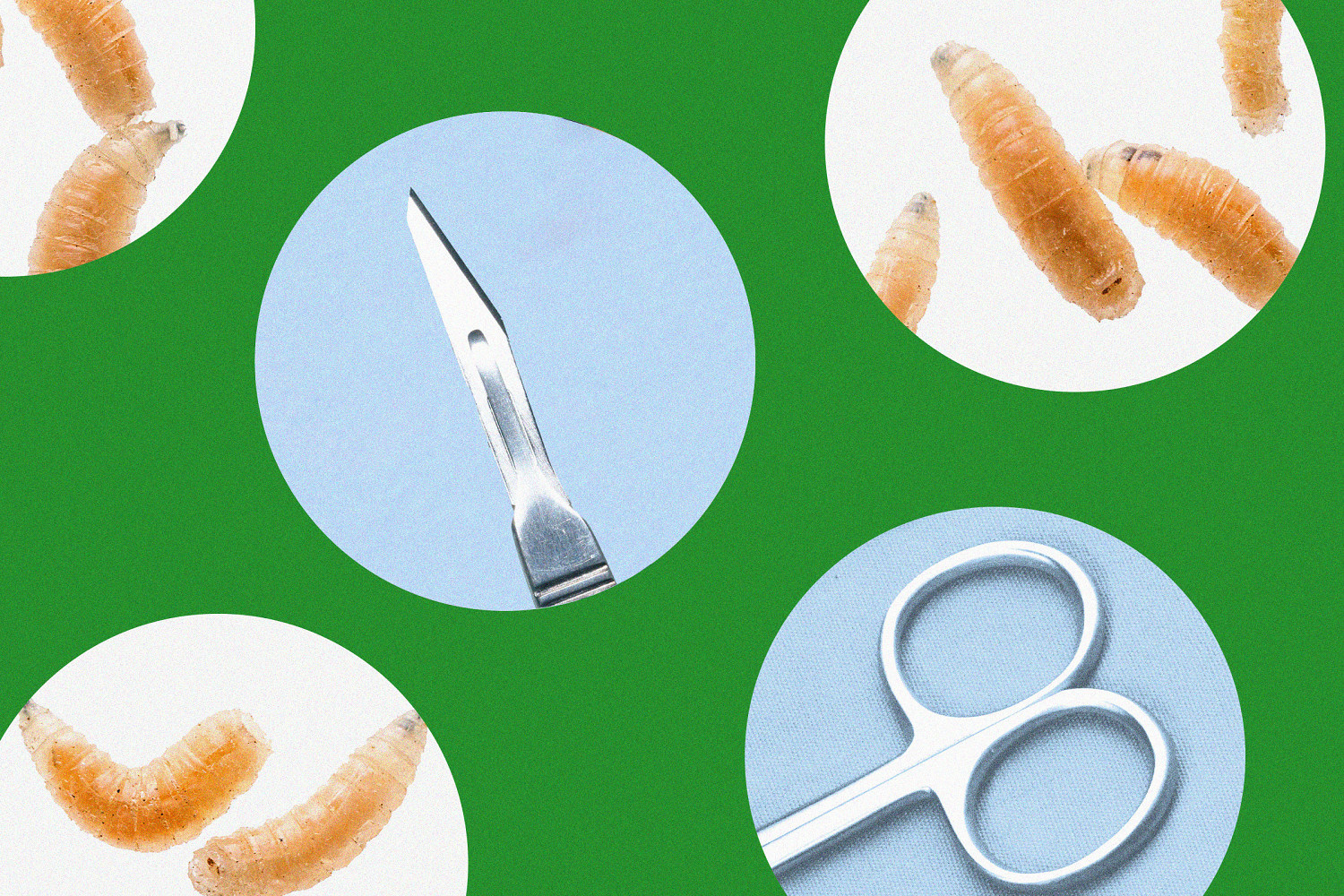
Chronic, non-healing wounds present a severe clinical bottleneck in modern healthcare, often resulting in prolonged hospitalization, systemic infection, or amputation. When conventional surgical debridement is contraindicated due to patient comorbidity, tissue location, or poor vascularity, Maggot Debridement Therapy (MDT) serves as a controlled, biological alternative. This modality relies on the introduction of sterile larvae of the blowfly, Lucilia sericata, into necrotic tissue. To evaluate MDT as a viable alternative to surgical intervention, we must analyze its efficacy through three distinct biological mechanisms: biochemical debridement, antimicrobial action, and the physical stimulation of granulation tissue.
The Tri-Centric Mechanism of Action
The clinical utility of Lucilia sericata is not driven by random consumption of tissue, but by a highly specialized, localized metabolic process. The larvae lack masticatory structures; instead, they rely on extracorporeal digestion. Understanding this process requires breaking down the therapy into its three functional pillars. For another perspective, see: this related article.
1. Enzymatic Debridement and the Cost Function of Necrotic Tissue
Larvae secrete a complex mixture of proteolytic enzymes, primarily matrix metalloproteinases (MMPs), serine proteases (trypsin-like and chymotrypsin-like), and aspartyl proteases. These secretions selectively degrade the extracellular matrix of necrotic tissue while leaving healthy, viable tissue intact.
Healthy human tissue possesses endogenous protease inhibitors (such as alpha-1-antitrypsin and alpha-2-macroglobulin) that neutralize these larval enzymes upon contact. Necrotic tissue lacks these active defense mechanisms. The biochemical cost function of the wound changes as liquefied necrotic debris is ingested by the larvae, effectively removing the biological substrate that fuels bacterial proliferation without inflicting the mechanical trauma associated with a scalpel. Similar insight on the subject has been shared by Medical News Today.
2. Antimicrobial Secretions and Biofilm Disruption
Wound chronicity is largely driven by biofilms—complex, polymicrobial matrices that shield bacteria from both the patient’s immune response and systemic antibiotic therapies. MDT disrupts this matrix through two distinct pathways:
- Chemical Neutralization: Larval secretions contain antimicrobial peptides (AMPs) such as lucifensin, alongside ammonium carbonate and allantoin. These compounds alter the local wound pH, shifting it from an acidic, infection-friendly state to an alkaline environment (pH 8.0–8.5) that inhibits the growth of common pathogens like Staphylococcus aureus and Pseudomonas aeruginosa.
- Mechanical Ingestion: As larvae browse the wound bed, they physically ingest bacteria trapped within the biofilm. The internal digestive tract of the larva acts as a sterilization chamber, destroying pathogens via internal enzymatic action and highly acidic midgut conditions.
3. Cellular Upshift and Granulation Stimulation
Beyond removal and disinfection, the wound bed requires a structural rebuild. Larval secretions contain urea, allantoin, and phenylacetic acid, which actively stimulate human fibroblast proliferation and migration. Furthermore, the physical movement of the larvae across the wound bed creates micro-mechanical forces. This low-level mechanical stimulation triggers the release of civil cytokines and growth factors, accelerating angiogenesis—the formation of new blood vessels—which transforms a stagnant, chronic wound into an active, healing environment.
Operational Logistics and Containment Engineering
Implementing MDT requires a strict adherence to containment protocols to manage patient compliance, exudate volume, and larval escape vectors. The application is bifurcated into two primary systems: the free-range technique and the contained (bagged) dressing system.
Free-Range vs. Contained Systems
The choice between these two delivery models dictates the operational efficiency and patient tolerance of the treatment.
The free-range system involves placing sterile larvae directly onto the wound bed and securing them with a breathable, fine-meshed netting anchored by hydrocolloid barriers. This configuration maximizes the physical mobility of the larvae, allowing them to access deep undermining, tracking tunnels, and irregular wound contours. The primary limitation of this approach is the requirement for intensive nursing oversight to prevent larval migration outside the treatment zone, alongside a higher incidence of localized discomfort reported by the patient due to direct crawling sensations.
The contained system utilizes a pre-manufactured, sealed polyvinyl alcohol or polyester pouch containing the larvae, cushioned by a porous spacer. The larval secretions freely pass through the porous membrane to liquefy necrotic tissue, and the liquefied matrix is sucked back into the pouch. While this system significantly lowers the psychological barrier for the patient and simplifies dressing changes for clinical staff, it reduces debridement velocity. The physical barrier restricts larval movement, preventing them from entering deep fissures or tunneling wounds effectively.
Dosing and Kinetic Limits
The standard clinical calculation for larval deployment is 5 to 10 larvae per square centimeter of wound surface area. A typical cycle lasts between 48 and 72 hours.
During this window, the larvae undergo a massive metabolic expansion, increasing their body mass up to 50-fold. This rapid growth creates a structural ceiling: once the larvae reach their third instar stage, they cease feeding and prepare to pupate. At this precise crossroad, the dressing must be removed. Leaving the larvae in place past the 72-hour mark yields zero additional debridement utility and increases the risk of larval escape or tissue maceration due to accumulated metabolic waste.
Comparative Matrix: MDT vs. Standard Care Modalities
To determine where MDT fits within a clinical protocol, it must be weighed against alternative debridement strategies across specific vectors: speed, tissue selectivity, pain profile, and systemic strain.
| Vector | Surgical Debridement | Maggot Debridement Therapy | Autolytic Debridement (Hydrogels) |
|---|---|---|---|
| Selectivity | Low to Moderate (Sacrifices viable margin) | Absolute (Biochemically targeted) | High (Enzyme reliant) |
| Velocity | Immediate (Minutes) | Rapid (48–72 Hours) | Very Slow (Weeks to Months) |
| Anesthesia Required | General or Local Required | None | None |
| Systemic Strain | High (Risk of blood loss, cardiac stress) | Low (Localized application) | Low (Passive process) |
| Cost Profile | High (Operating room fees, surgeon time) | Moderate (Specialized biological materials) | Low (Standard dressing consumables) |
Surgical debridement offers unmatched immediacy, making it the non-negotiable choice for acute, life-threatening infections like necrotizing fasciitis. However, surgery is an indiscriminate process; surgeons often must excise a margin of healthy, viable tissue to guarantee complete removal of the necrosis.
MDT serves as an optimization strategy for patients who cannot tolerate the physiological stress of surgery—such as those suffering from advanced peripheral arterial disease, severe cardiovascular insufficiency, or brittle diabetes. It achieves a level of tissue selectivity that no human hand can replicate, removing non-viable material atom by atom while leaving delicate microvascular structures intact.
Clinical Boundaries, Risks, and Contraindications
MDT is a highly specialized tool, not a panacea. Successful implementation requires recognizing its strict boundaries to avoid catastrophic tissue failure.
Absolute Contraindications
Treating certain anatomical structures with MDT introduces unacceptable risks. Larvae cannot differentiate between necrotic tissue and the compromised, ischemic walls of major blood vessels. Applying larvae to wounds in close proximity to large arteries or veins—such as an exposed femoral artery in a groin wound—creates a severe risk of lethal hemorrhage.
Similarly, wounds that connect directly to internal body cavities, organ systems, or deep synovial joint spaces are entirely excluded from this therapy. Uncontrolled larval migration into these sterile environments can cause deep-seated infections or mechanical damage to vital organs.
Relative Contraindications and Complications
- Ischemic Thresholds: For MDT to stimulate granulation tissue, the wound bed must possess a baseline level of arterial perfusion. In cases of severe peripheral arterial disease where the Ankle-Brachial Index (ABI) falls below 0.5, removing the necrotic tissue will not induce healing; it will simply expose ischemic bone or tendon that cannot regenerate due to lack of blood supply. Revascularization must precede biotherapy.
- Localized Pain Management: While the debridement process itself is un-innervated, the resulting drop in local pH and the physical volume increase of the larvae within a confined space can compress local nerve endings. This typically manifests during the final 24 hours of a cycle. Managing this requires a proactive escalation of non-opiate analgesics and a willingness to terminate the cycle early if pain thresholds are breached.
- Psychological and Cultural Barriers: The "revulsion factor" remains a significant bottleneck to widespread adoption. Patients and healthcare providers frequently exhibit deep-seated resistance to biological therapies. Mitigating this requires reframing the conversation around objective metrics: reducing total healing time, avoiding operating rooms, and lowering the risk of amputation.
Deployment Blueprint
To integrate Maggot Debridement Therapy into a chronic wound care protocol, clinical teams should execute the following deployment sequence:
- Hemodynamic Verification: Confirm adequate macro-vascular perfusion via arterial Doppler ultrasound or ensure the patient has an ABI of $\ge 0.6$ before ordering biological materials.
- Periwound Protection: Apply a high-viscosity zinc oxide or cyanoacrylate skin barrier to the healthy skin surrounding the wound perimeter. This protects intact tissue from maceration caused by the highly proteolytic larval runoff.
- Dressing Selection: Deploy free-range systems for deep, irregular tracking wounds to leverage larval mobility. Utilize contained pouch systems for flat, superficial ulcers on highly sensitive patients to maximize compliance.
- Exudate Management: Layer a highly absorbent, non-occlusive secondary dressing over the containment mesh. The larvae require oxygen to survive; sealing them beneath hydrocolloids or film dressings will cause asphyxiation and treatment failure.
- Cycle Termination: Remove the dressing strictly between 48 and 72 hours. Flush the wound bed with sterile saline to remove remaining larvae, dispose of all biohazardous materials according to regional infectious waste protocols, and reassess the wound bed for sequential cycles or immediate transition to standard moist wound-healing therapies.